My main areas of research are medical image processing and cancer imaging. In particular, my research focuses on: breast cancer, liver cancer, machine learning, and 3D visualization and printing.
Artificial intelligence for breast cancer imaging
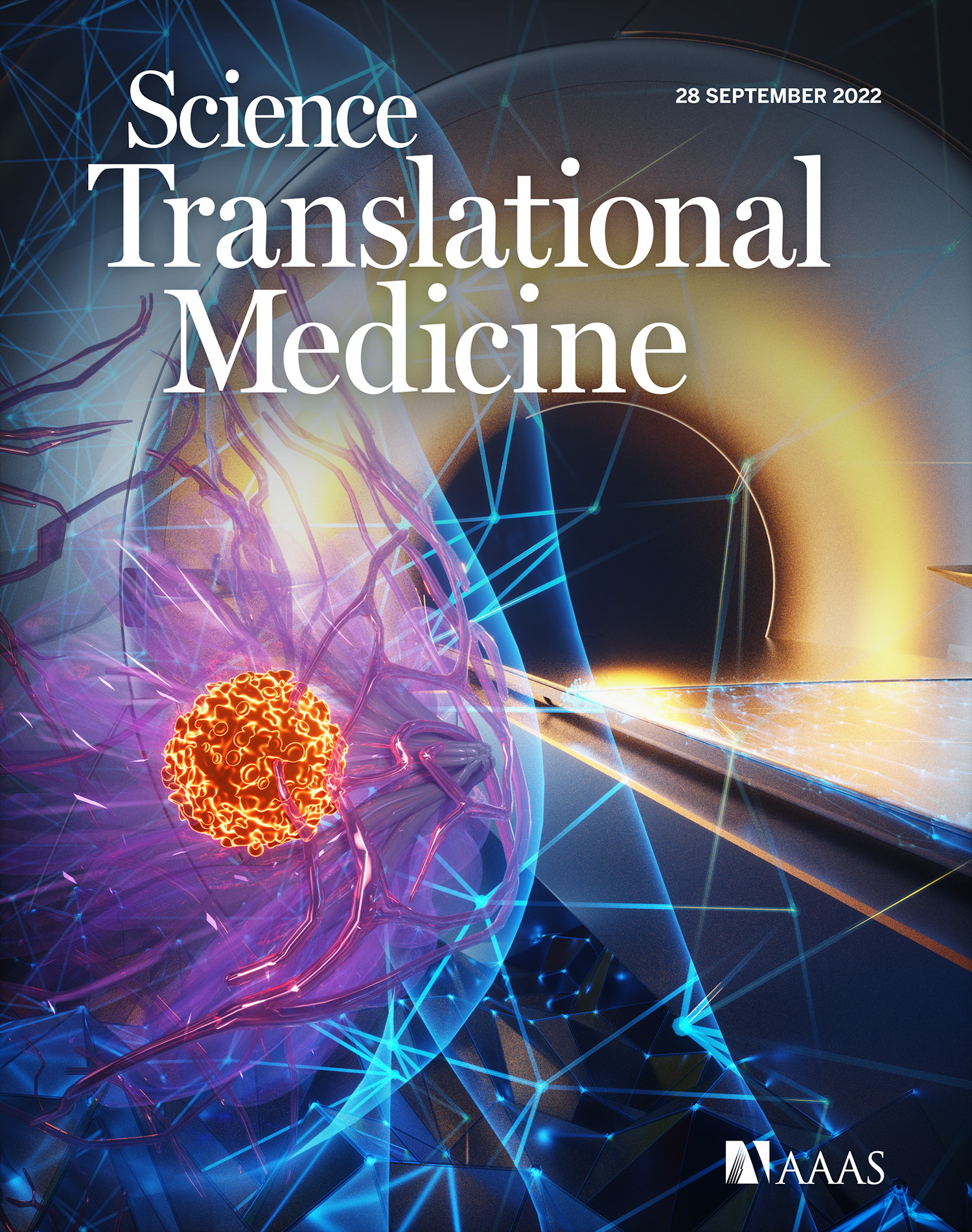
In years 2020-2022, during my postdoc at NYU, my work was focused on the AI in multi-modal breast cancer imaging (mammography, tomosynthesis, ultrasound, MRI). I've been a project leader of breast cancer classification in MRI with convnets. Results of the breast MRI project were published in Science Translational Medicine in September 2022, as well as featured on the journal's cover. Our robust, retrospective analysis showed that our system could reduce the number of unnecessary biopsies by up to 20 percent. We evaluated our findings on multiple, international data sets, including a data set from my Alma Mater -- Jagiellonian University in Kraków, Poland.
With other NYU team members, I participated and won the Digital Breast Tomosynthesis Lesion Detection Challenge, organized by the SPIE, the American Association of Physicists in Medicine (AAPM), the National Cancer Institute (NCI), and Duke AI in Radiology (DAIR). We summarized our results in a Nature Machine Intelligence paper. You can also find a story about this challenge on CAI2R website or a video of the winning entry, presented by project leader, Jungkyu Park.
My work is highly focused on clinical outcomes and translation of results to the clinical side - statistical versus clinical significance, integration with clinical workflow, societal impact. I work closely with breast imaging radiologists, including renowned Dr. Linda Moy, Dr. Laura Heacock and Dr. Beatriu Reig.
I managed and wrangled multi-million-item datasets of multimodal medical images and EHR for AI system development. This dataset has been used in our lab's work in all modalities, including mammography, tomosynthesis, ultrasound and MRI.
In August 2021 we published a meta-repository which combines international mammography datasets with state-of-the-art AI models for breast cancer classification. This is an open-source project and the code is publicly available on GitHub.
3D printing for liver cancer surgery
In years 2016-2020, during my MD/PhD program, I worked with a team of oncological surgeons at Jagiellonian University in Kraków to develop 3D printed anatomical models for preoperative planning. I invented a low-cost, reproducible method of 3D printing full-sized, multicolor liver models for planning laparoscopic liver resections. In my publications, I validated the accuracy of those models and later ran a prospective pilot study to measure clinical feasibility and importance. As a result of those papers, I've confirmed that 3D printed liver models are accurate enough to be used in the clinical practice, and their use in preoperative planning can make meaningful changes in the surgery plan in every 4th patient. My work also made an impact on developing RSNA's guidelines for clinical 3D printing.
My method of 3D printing liver models has been covered in Polish and international media:
Education
I have recorded tutorials on YouTube for 3D Slicer, an open-source software designed for medical image processing, that have been watched over 50,000 times. I have trained tens of researchers worldwide in using 3D Slicer for image analysis, including medical segmentation and more. Occasionally, I am a speaker during lectures, workshops, conferences etc. on medical imaging and new technologies in medicine, such as 3D printing or artificial intelligence. During my postdoc, I supervised work of three MS students at Harvard & NYU.